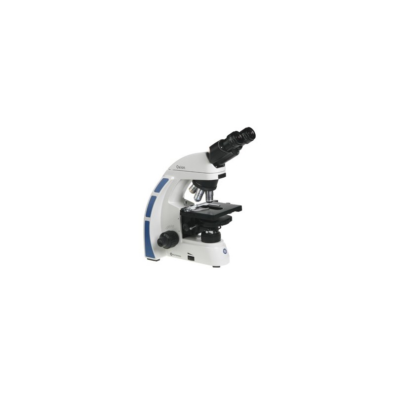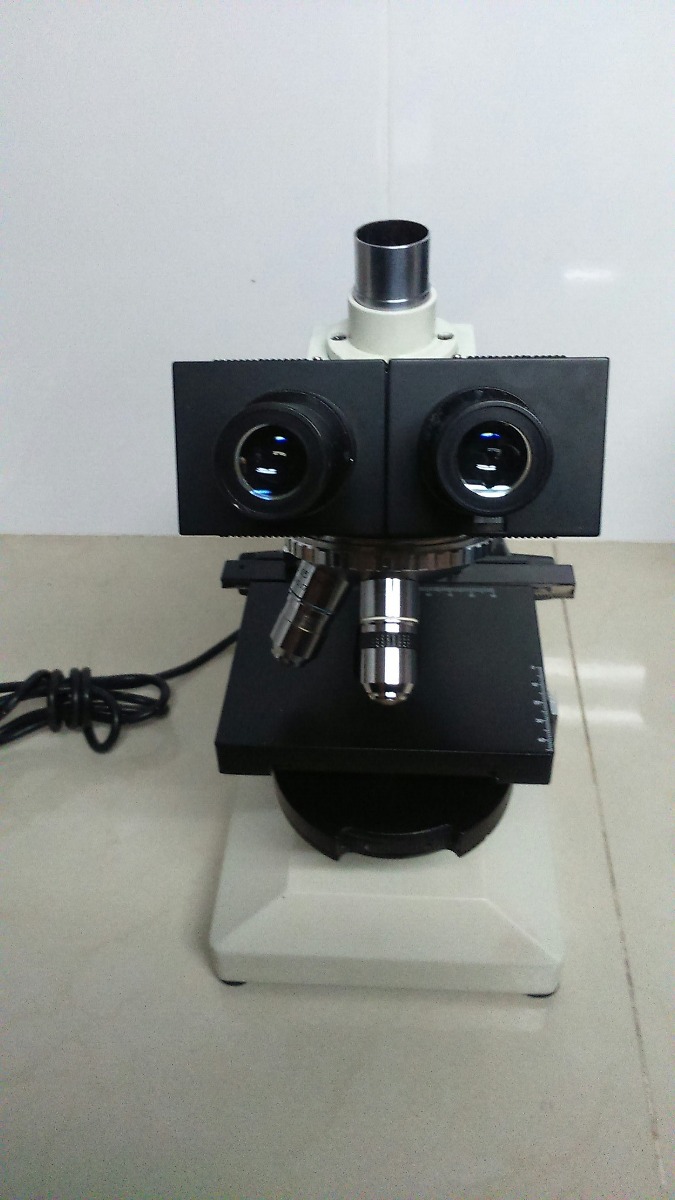

The optical system of the phase-contrast microscope differs from the ordinary (light) microscope only in the addition of an annular diaphragm placed under the stage to illuminate the object with a narrow light cone and a diffraction plate mounted on the lens.

Thus, in the representation of Figure 1, the ray passing through part B is delayed 1/4 wavelength (l/4) and emerges out of phase, with respect to the transmission shown in part A. If the difference in refractive indexes is small, the magnitude of the induced phase change is also very small and measured in terms of wavelengths (l). In this specific case, it takes longer to pass light through fraction B, whose refractive index is higher (n2) and therefore the lightning wave is delayed in speed respect to which it is able to traverse the portion A, whose refractive index is lower. Variations in thickness and refractive indexes are capable of producing a difference in the optical course of light transmitted by the two regions. Figure 1 shows the schematic representation in which two adjacent regions of the same cell, A and B, of a different thickness, t1 and t2 and with different refractive indexes, n1 and n2, are crossed by a luminous ray. However, it finds in its path regions of different refractive indexes, and different thicknesses and/or densities, which is able to alter their speed and direction. The light is transmitted through living cells. Living cells are transparent to visible light, so light rays pass through without loss of intensity.

So, these optical instruments help to solve the problem. Phase-contrast microscopy and the interference microscope give us the possibility to observe living cells without any preparation, since when performing other procedures there is the possibility that some of its components may be lost or distorted by methods such as fixation, coloring, freezing, and others. 1953 Nobel Prize in Physics awarded to Zernike.1952 Nomarski : Invents and patents the Interferencel contrast system that bears his name.1932 Frits Zernike : Invents phase-contrast microscope.1930 Labedeff : Designs first interference contrast microscope.Both types of optical microscopes are commonly used to visualize living cells. The phase-contrast microscope and the interference phase-contrast microscope take advantage of the interference effects that occur when these two groups of waves are combined, thus creating an image of the cell structure. When light passes through a living cell, the phase of the light wave varies in relation to the refractive index of the cell: the light that passes through a relatively thick or dense area of the cell, such as the nucleus, is delayed and its phase is displaced correspondingly to that of light that has passed through an adjacent, finer cytoplasm region. Microscopes with special optical systems are useful for this purpose. The only solution to this problem is to examine the cells while they are still alive, without any fixation or freezing. The possibility that some cell components may be lost or distorted during sample preparation has not ceased to worry microscopy specialists. Live cells can be clearly observed under a phase-contrast or interference phase-contrast microscope.


 0 kommentar(er)
